The heart is a central organ enabling blood circulation, located in the thoracic cavity. Its intricate structure includes chambers, valves, and vessels like the aorta, ensuring efficient oxygen delivery.
Overview of the Heart’s Structure and Function
The heart is a muscular organ acting as a pump, located in the thoracic cavity. Its structure includes four chambers, valves, and blood vessels like the aorta. The heart’s primary function is to circulate blood, supplying oxygen and nutrients to tissues while removing waste. This efficient system ensures survival, making the heart a vital organ for overall health. Understanding its structure and function is essential for medical diagnostics and treatments.
Importance of Understanding Heart Anatomy
Understanding heart anatomy is crucial for diagnosing and treating cardiovascular diseases. It aids in identifying structural defects, such as faulty valves or abnormal chambers, and guides surgical interventions. Knowledge of heart structure helps develop life-saving treatments and improves patient outcomes. Studying anatomy also fosters a deeper appreciation for cardiovascular health and prevents complications. This understanding is vital for advancing medical research and improving heart disease management.
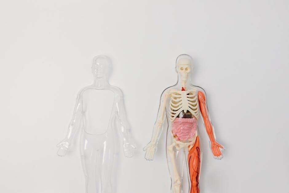
Chambers of the Heart
The heart contains four chambers: right atrium, left atrium, right ventricle, and left ventricle. They work together to receive and pump blood efficiently throughout the body.
Right Atrium and Left Atrium
The right atrium receives deoxygenated blood from the body via the superior and inferior vena cava, while the left atrium receives oxygenated blood from the lungs via the pulmonary veins. Both atria act as reservoirs, ensuring a steady blood flow into the ventricles. Their thin walls allow for passive filling, playing a crucial role in the heart’s continuous pumping cycle and overall circulatory efficiency.
Right Ventricle and Left Ventricle
The right ventricle pumps deoxygenated blood through the pulmonary valve to the lungs, while the left ventricle pushes oxygenated blood through the aortic valve to the body. The left ventricle has thicker walls due to higher pressure demands. Both ventricles play vital roles in maintaining systemic and pulmonary circulation, ensuring efficient oxygenation and nutrient delivery to tissues throughout the body.

Layers of the Heart Wall
The heart wall consists of three layers: the pericardium, myocardium, and endocardium. Each layer performs distinct functions essential for cardiac structure and optimal blood circulation.
Pericardium
The pericardium is the outermost layer of the heart wall, functioning as a protective fibroserous sac. It encloses the heart and the roots of the great vessels, providing structural support and safeguarding against external shocks. The pericardium consists of the fibrous pericardium and the serous pericardium, with the latter producing pericardial fluid to lubricate and reduce friction during heart contractions, ensuring smooth cardiac function and overall cardiovascular health.
Myocardium
The myocardium is the thick, muscular middle layer of the heart wall, primarily composed of cardiac muscle cells. It is responsible for the heart’s contractile function, enabling the pumping of blood through the chambers and into the circulatory system. The myocardium contains bundles of muscle fibers that coordinate contractions, ensuring efficient blood circulation. It is richly supplied with blood by coronary arteries, which are essential for maintaining its functionality and overall cardiac health.
Endocardium
The endocardium is the innermost layer of the heart wall, lining the heart chambers and valves. It is a thin, smooth layer of connective tissue covered by endothelial cells, preventing blood clotting and ensuring smooth blood flow. The endocardium plays a critical role in maintaining the heart’s internal environment and regulating blood circulation. Damage to this layer can lead to conditions such as endocarditis, highlighting its importance in cardiac health;

Valves of the Heart
The heart contains four valves ensuring blood flows in one direction. These valves prevent backflow, enabling efficient circulation of oxygenated and deoxygenated blood throughout the body.
Types of Heart Valves
The heart features four main valves: the tricuspid, pulmonary, mitral (bicuspid), and aortic valves. Each valve ensures unidirectional blood flow, preventing backflow. The tricuspid valve is between the right atrium and ventricle, while the pulmonary valve connects the right ventricle to the pulmonary artery. The mitral valve lies between the left atrium and ventricle, and the aortic valve links the left ventricle to the aorta, facilitating blood distribution throughout the body.
Function and Significance of Each Valve
Each heart valve ensures unidirectional blood flow, maintaining cardiac efficiency. The tricuspid valve controls blood movement from the right atrium to the ventricle, while the pulmonary valve regulates flow to the lungs. The mitral valve manages blood flow from the left atrium to the ventricle, and the aortic valve directs oxygenated blood to the body via the aorta. Proper valve function is crucial for preventing backflow and maintaining optimal circulation.

Blood Flow Through the Heart
The heart facilitates blood circulation through pulmonary and systemic pathways, directing deoxygenated blood to the lungs and oxygenated blood to the body, ensuring efficient delivery of oxygen.
Pulmonary Circulation
Pulmonary circulation begins in the right ventricle, where deoxygenated blood flows through the pulmonary artery to the lungs. Oxygen is absorbed into the blood, which then returns via pulmonary veins to the left atrium, completing the cycle. This pathway ensures oxygenation of blood, a critical process for cellular respiration and overall bodily function, highlighting the heart’s role in maintaining life-sustaining oxygen delivery.
Systemic Circulation
Systemic circulation transports oxygenated blood from the left ventricle through the aorta to the body’s tissues and organs. The aorta branches into arteries, distributing oxygen and nutrients. Deoxygenated blood returns via veins to the right atrium, completing the cycle. This system ensures vital organs receive the necessary resources for function, with the coronary arteries specifically supplying the heart itself, maintaining its operation and overall bodily health.
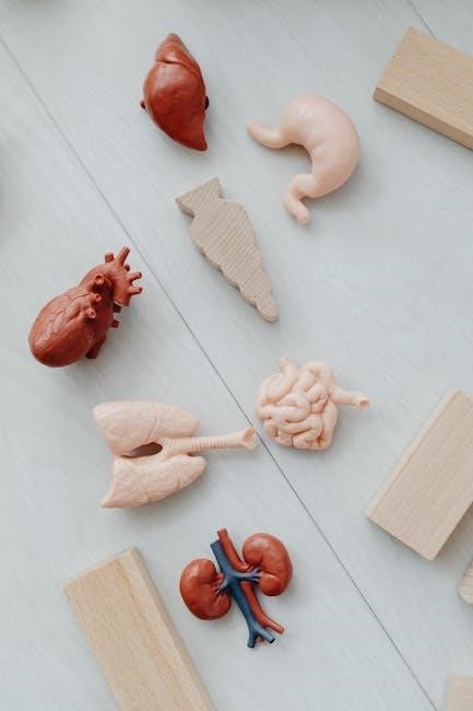
The Electrical System of the Heart
The heart’s electrical system regulates its rhythmic contractions, starting with the sinoatrial node, the natural pacemaker. It ensures consistent heartbeat timing, essential for proper blood circulation and overall health.
Sinoatrial Node and Atrioventricular Node
The sinoatrial node, located in the right atrium, acts as the heart’s natural pacemaker, initiating electrical impulses for contractions. The atrioventricular node relays these signals to the ventricles, ensuring synchronized heartbeat timing. Together, they regulate the heart’s rhythmic contractions, maintaining proper blood flow and overall cardiac function.
Bundle of His and Purkinje Fibers
The Bundle of His transmits electrical impulses from the atrioventricular node to the ventricles, ensuring coordinated contraction. Purkinje fibers, branching from the Bundle of His, spread the signal across the ventricular walls. This specialized conduction system enables rapid, synchronized contractions, maintaining efficient blood pumping and proper heart rhythm.

Clinical Relevance of Heart Anatomy
Understanding heart anatomy aids in diagnosing and treating cardiovascular diseases, improving patient outcomes. It helps identify structural abnormalities and guides interventions like surgeries or catheterizations effectively.
Congenital Heart Defects
Congenital heart defects are structural abnormalities in the heart present at birth, often affecting chambers, valves, or blood vessels; Common defects include septal defects and valve malformations. These defects can disrupt normal blood flow, leading to symptoms like cyanosis, rapid breathing, or poor feeding in infants. Early diagnosis, often through echocardiography, is critical for timely intervention. Treatments range from monitoring to surgical or catheter-based procedures, improving long-term outcomes for affected individuals.
Acquired Heart Diseases
Acquired heart diseases develop over time and are not present at birth. Conditions like coronary artery disease, hypertension, and heart failure are common. These diseases often result from lifestyle factors, age-related changes, or infections. Symptoms may include chest pain, breathlessness, or fatigue; Early diagnosis through imaging and tests is crucial. Treatments vary, ranging from medications to surgery, and focus on managing symptoms and improving heart function to enhance quality of life.

Diagnostic Imaging of the Heart
Diagnostic imaging of the heart involves techniques like X-rays, CT scans, and MRIs to examine heart structures and detect abnormalities, aiding in accurate diagnoses and treatments.
Echocardiography
Echocardiography is a non-invasive imaging technique that uses ultrasound waves to produce detailed images of the heart’s structure and function. It is widely used to assess heart valves, chambers, and blood flow.
This method provides real-time visualization of cardiac activity, helping diagnose conditions such as valve disorders, heart disease, and abnormalities in heart function. Its safety and effectiveness make it a crucial tool in cardiovascular diagnostics.
Cardiac MRI
Cardiac MRI uses magnetic fields and radio waves to create high-resolution images of the heart’s anatomy and function. It provides detailed views of heart structures, such as chambers, valves, and blood vessels, aiding in the diagnosis of various cardiac conditions.
This non-invasive technique is particularly effective for assessing heart tissue viability, detecting abnormalities, and evaluating the severity of heart disease, offering precise insights for treatment planning.
Understanding heart anatomy is crucial for appreciating its function and addressing cardiac health. Study of its structure aids in preventing and managing heart diseases effectively.
The heart, a muscular organ, consists of four chambers: two atria and two ventricles. It pumps blood through pulmonary and systemic circulation, regulated by valves and an electrical system. The pericardium, myocardium, and endocardium form its layers. Understanding its anatomy aids in diagnosing defects and diseases, emphasizing the importance of detailed study for effective cardiac care and treatment.
Importance of Continued Study
Continued study of heart anatomy is crucial for advancing medical knowledge and improving patient care. It enhances understanding of cardiac structures, aiding in early diagnosis and treatment of heart diseases. Ongoing research into the heart’s anatomy supports the development of new therapies, surgical techniques, and diagnostic tools, ultimately improving outcomes for individuals with cardiac conditions and advancing cardiovascular medicine.


